What Exactly Is Posterior Calcaneal Spur

Posterior Calcaneal Spur

Overview

The heel bone is the largest bone in the foot and absorbs the most shock and pressure. A heel spur develops as an abnormal growth of the heel bone. Most commonly, calcium deposits form when the plantar fascia pulls away from the heel area, causing a bony protrusion, or heel spur to develop. While bone spurs can develop in other locations such as the rear of the heel bone, this is the most common location for development. The plantar fascia is a broad band of fibrous tissue located along the bottom surface of the foot that runs from the heel to the forefoot. Heel spurs can cause extreme pain in the rearfoot, especially while standing or walking.

Causes

Heel spurs are bony outgrowths positioned where the plantar fascia tissue attaches to the heel bone (the calcaneus). Heel spurs seldom cause pain. It is the inflamed tissue surrounding the spur that causes the pain. The Latin meaning of Plantar Fasciitis is, ?Inflammation of Plantar Fascia.? The plantar fascia is a long, thick and very tough band of tissue beneath your foot that provides arch support. It also connects your toes to your heel bone. Each time you take a step, the arch slightly flattens to absorb impact. This band of tissue is normally quite strong and flexible but unfortunately, circumstances such as undue stress, being overweight, getting older or having irregularities in your foot dynamics can lead to unnatural stretching and micro-tearing of the plantar fascia. This causes pain and swelling at the location where the plantar fascia attaches to the heel bone. As the fascia continually pulls at the heel bone, the constant irritation eventually creates a bony growth on the heel. This is called a heel spur.

Heel Spur

Symptoms

Heel spur and plantar fasciitis pain usually begins in the bottom of the heel, and frequently radiates into the arch. At times, however, the pain may be felt only in the arch. The pain is most intense when first standing, after any period of rest. Most people with this problem experience their greatest pain in the morning, with the first few steps after sleeping. After several minutes of walking, the pain usually becomes less intense and may disappear completely, only to return later with prolonged walking or standing. If a nerve is irritated due to the swollen plantar fascia, this pain may radiate into the ankle. In the early stages of Heel Spurs and Plantar Fasciitis, the pain will usually subside quickly with getting off of the foot and resting. As the disease progresses, it may take longer periods of time for the pain to subside.

Diagnosis

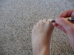
A Heel Spur diagnosis is made when an X-ray shows a hook of bone protruding from the bottom of the foot at the point where the plantar fascia is attached to the heel bone. The plantar fascia is the thick, connective tissue that runs from the calcaneus (heel bone) to the ball of the foot. This strong and tight tissue helps maintain the arch of the foot. It is also one of the major transmitters of weight across the foot as you walk or run. In other words, tremendous stress is placed on the plantar fascia.

Non Surgical Treatment

Common and effective treatments for Heel Spurs include: Stretching exercises, changing to specific shoes, taping or strapping to rest stressed muscles and tendons, custom orthotic devices and physiotherapy. There are many things you can do to treat heel spurs. You should stretch the muscles and ligaments around the area regularly and ensure you are wearing the right footwear for your feet. There are also tapes and straps that you can apply to the muscles and tendons around the area. For more severe cases, custom orthotics may be the way to go along with aggressive physiotherapy. To treat the pain, over the counter NSAIDs (anti-inflammatory medications) is recommended, but use with caution as prolonged use can lead to the development of ulcers. It is therefore best to apply a topical treatment such as Zax?s Original Heelspur Cream, which contains natural ingredients proven to reduce pain and inflammation. More severe forms of the condition may require corticosteroid injections or surgical procedures, but these are very rare cases. Still, should pain become worse and persist, you should consult with your doctor.

Surgical Treatment

Approximately 2% of people with painful heel spurs need surgery, meaning that 98 out of 100 people do well with the non-surgical treatments previously described. However, these treatments can sometimes be rather long and drawn out, and may become considerably expensive. Surgery should be considered when conservative treatment is unable to control and prevent the pain. If the pain goes away for a while, and continues to come back off and on, despite conservative treatments, surgery should be considered. If the pain really never goes away, but reaches a plateau, beyond which it does not improve despite conservative treatments, surgery should be considered. If the pain requires three or more injections of “cortisone” into the heel within a twelve month period, surgery should be considered.

Bursitis Of The Feet Surgical Treatment

Overview

Bursitis is the inflammation of a bursa within the body. A bursa is a fluid-filled sac that cushions some of the major joints, allowing bones, gliding muscles, and tissues to exist together without harming each other. It aids in reducing the friction between bones and the soft tissues above it. Though there are bursae all over the body, the main ones are located in the knee, elbow, shoulder, hip, and heel. These are also the ones that are the most often injured. Still, however, when you hear the word ?bursitis?, the person is usually referring to their shoulder, but it could also refer to any of the preceding areas–such as the second most common one, the hip.

Causes

Bursitis occurs when the bursae become irritated or infected, often causing pain on movement. When infection is involved, medical intervention is necessary to fight the underlying infection and prevent it from spreading, when infection is not involved, prompt medical attention can prevent the condition from becoming worse over time.

Symptoms

Pain in the heel, especially with walking, running, or when the area is touched. Pain may get worse when rising on the toes (standing on tiptoes). Red, warm skin over the back of the heel.

Diagnosis

On physical examination, patients have tenderness at the site of the inflamed bursa. If the bursa is superficial, physical examination findings are significant for localized tenderness, warmth, edema, and erythema of the skin. Reduced active range of motion with preserved passive range of motion is suggestive of bursitis, but the differential diagnosis includes tendinitis and muscle injury. A decrease in both active and passive range of motion is more suggestive of other musculoskeletal disorders. In patients with chronic bursitis, the affected limb may show disuse atrophy and weakness. Tendons may also be weakened and tender.

Non Surgical Treatment

Caregivers may give you special shoe inserts with a cutout around the tender area. You may also be told to wear shoes with a reinforced heel counter. This will give better heel control. You may need other shoe inserts (wedges) to raise your heel so it does not press against the back of the shoe. You may also wear shoes that are open in the back, such as sandals that have no strap across the heel. You may use ibuprofen (eye-bu-PROH-fen) and acetaminophen (a-seet-a-MIN-oh-fen) medicine for your pain. These may be bought over-the-counter at drug or grocery stores. Do not take ibuprofen if you are allergic to aspirin. You may be given shots of medicine called steroids (STER-oids) to decrease inflammation. Caregivers may add local anesthesia (an-es-THEE-zah) to the steroids. This medicine helps decrease bursitis pain. Because these shots decrease swelling and pain, you may feel like your ankle is healed and that you can return to heavy exercise. It is important to not exercise until your caregiver says it is OK. You could make the bursitis worse if you exercise too soon. You may need surgery to remove the bursa or part of your ankle bone. Surgery is usually not necessary unless the bursitis is very bad and does not heal with other treatments. Your caregiver may want you to go to physical (FIZ-i-kal) therapy (THER-ah-pee). Physical therapists may use ultrasound to increase blood flow to the injured area. Caregivers may use massage to stretch the tissue and bring heat to the injury to increase blood flow. These and other treatments may help the bursitis heal faster. Exercises to stretch your Achilles tendon and make it stronger will be started after the bursitis has healed. You may gradually increase the amount of weight you put on your foot when caregivers say it is OK. You may be told to stop exercising if you feel any pain.

Surgical Treatment

Surgery is rarely done strictly for treatment of a bursitis. If any underlying cause is the reason, this may be addressed surgically. During surgery for other conditions, a bursa may be seen and removed surgically.

Hammer Toe Treatment Surgery

Hammer ToeOverview

hammertoe is a condition which causes one or more of the smaller toes to become bent upwards. The toe can be straightened but if ignored may become a permanent deformity. Each of the 4 smaller toes consist of 3 bones called phalanges, forming two interphalangeal joints. The toe bends at the proximal or first interphalangeal joint. Initially it can be straightened, but if left untreated, this can become a permanent deformity.

Causes

More often than not, wearing shoes that do not fit a person well for too long may actually cause hammer toes. Wearing shoes that are too narrow or too tight for the person for extended periods of time may eventually take a toll on the person’s feet. The same is true for women who like wearing high-heeled shoes with narrow toe boxes.

HammertoeSymptoms

The middle joint of the toe is bent. The end part of the toe bends down into a claw-like deformity. At first, you may be able to move and straighten the toe. Over time, you will no longer be able to move the toe. It will be painful. A corn often forms on the top of the toe. A callus is found on the sole of the foot. Walking or hammertoe wearing shoes can be painful.

Diagnosis

The treatment options vary with the type and severity of each hammer toe, although identifying the deformity early in its development is important to avoid surgery. Your podiatric physician will examine and X-ray the affected area and recommend a treatment plan specific to your condition.

Non Surgical Treatment

Conservative treatment starts with new shoes that have soft, roomy toe boxes. Shoes should be one-half inch longer than your longest toe. (Note: For many people, the second toe is longer than the big toe.) Avoid wearing tight, narrow, high-heeled shoes. You may also be able to find a shoe with a deep toe box that accommodates the hammer toe. Or, a shoe specialist (Pedorthist) may be able to stretch the toe box so that it bulges out around the toe. Sandals may help, as long as they do not pinch or rub other areas of the foot.

Surgical Treatment

If conservative treatments don’t help, your doctor may recommend surgery to release the tendon that’s preventing your toe from lying flat. In some cases, your doctor might also remove some pieces of bone to straighten your toe.

HammertoePrevention

Although these following preventative tips may be able to reverse a painful bunion or hammertoe deformity, they are more effective when applied to young people, and are less effective the longer a person has progressed with their bunion or hammertoe deformity. This is because the joints in our bodies get used to the positions they are most frequently held in, and our feet are no different, with our 12 to 15 hours a day in restrictive footwear, with tapering toeboxes, heel elevation, and toespring.

Hammer Toe Fusion Surgery

Hammer ToeOverview

If you sneak a peek at your feet and notice that your toes are crossed, bent, or just pointing at an odd angle, you probably suffer from a hammertoe. Toes that are scrunched up inside tight shoes or pressed against the toe box of the shoe can bend at the joints and stay that way – resulting in a hammertoe. A hammertoe is a contracture of the toe at one of the two joints in the toe. Due to the pull of the tendons, the joints become more rigid over time. The toe is bent up at the joint and does not straighten out.

Causes

Hammertoe commonly develops because of structural changes that take place over time in the muscles and tendons that bend the toes. People with certain medical conditions, such as diabetes, are at risk for developing hammertoe. It can be an inherited condition for some people. Other causes include trauma and wearing shoes that are too tight, narrow, or have high heels. The toe next to the big toe (second toe) is most frequently affected by hammertoe.

HammertoeSymptoms

For some people, a hammer toe is nothing more than an unsightly deformity that detracts from the appearance of the foot. However, discomfort may develop if a corn or callus develops on the end or top of the toe. If pressure and friction continue on the end or top of the toe, a painful ulcer may develop. Discomfort or pain can lead to difficulty walking.

Diagnosis

Hammer toes may be easily detected through observation. The malformation of the person’s toes begin as mild distortions, yet may worsen over time – especially if the factors causing the hammer toes are not eased or removed. If the condition is paid attention to early enough, the person’s toes may not be permanently damaged and may be treated without having to receive surgical intervention. If the person’s toes remain untreated for too long, however the muscles within the toes might stiffen even more and will require invasive procedures to correct the deformity.

Non Surgical Treatment

Your doctor may prescribe some toe exercises that you can do at home to stretch and strengthen the muscles. For example, you can gently stretch the toes manually. You can use your toes to pick things up off the floor. While you watch television or read, you can put a towel flat under your feet and use your toes to crumple it. Finally, your doctor may recommend that you use commercially available straps, cushions or nonmedicated corn pads to relieve symptoms. If you have diabetes, poor circulation or a lack of feeling in your feet, talk to your doctor before attempting any self-treatment.

Surgical Treatment

If pinning the toe is not required during the procedure, then the surgery could be preformed in the doctor’s office under a local anesthesia. Some patients prefer the comfort of sedation during the surgery and if this is the case or if a pin must be placed, then the surgery could be preformed in an outpatient surgery center.

HammertoePrevention

These tips may help you buy the right shoes. Buy shoes at the end of the day. Your feet are smaller in the morning and swell throughout the day. Don’t assume your shoe size hasn’t changed. As you age, your shoe hammertoe size may change, especially the width. Measure both feet and buy for the larger foot. Ask for just the right fit. A shoe repair store can stretch shoes in tight spots.

All You Want To Know Regarding Bunions

Overview
Bunions
A bunion is a prominence over the inside part of the foot where the big toe joins the rest of the foot. Pressure on the prominence from shoes causes pain and swelling due to inflammation. The bunion occurs when the foot bone connecting to the big toe (the first metatarsal) moves gradually towards the opposite foot. This is called hallux valgus deformity. This leads to the big toe being pushed towards the second toe (away from the opposite foot) so that the big toe points away from the other foot. Other problems can develop with a bunion. For example the second toe may overlap the big toe causing a cross-over toe deformity. With shoe pressure, corns and calluses develop.

Causes
Bunions most commonly affect women. Some studies report that bunion symptoms occur nearly 10 times more frequently in women. It has been suggested that tight-fitting shoes, especially high-heel and narrow-toed shoes, might increase the risk for bunion formation. Tight footwear certainly is a factor in precipitating the pain and swelling of bunions. Complaints of bunions are reported to be more prevalent in people who wear shoes than in barefoot people. Other risk factors for the development of bunions include abnormal formation of the bones of the foot at birth (congenital) and arthritic diseases such as rheumatoid arthritis. In some cases, repetitive stresses to the foot can lead to bunion formation. Bunions are common in ballet dancers.
SymptomsThe symptoms of a bunion include pain, swelling, and redness over the bony bump on the inside of the foot. It can become painful to walk, because the big toe bends every time you take a step. Shoes can become painful to wear, especially ones that are even a little bit tight. Usually, bunions become more painful as they get larger. In severe cases, you can develop arthritis in the big toe as a result of the bunion. However, a bunion that is not painful does not need surgical treatment, even a large one.

Diagnosis
A doctor can very often diagnose a bunion by looking at it. A foot x-ray can show an abnormal angle between the big toe and the foot. In some cases, arthritis may also be seen.

Non Surgical Treatment
The first step in the treatment of Bunions, Corns, and Calluses is determining what is causing the problem. Check with your family doctor or Podiatrist (foot doctor) to find the best solution for your ailment. You can reduce the risk of Bunions, Corns, and Calluses by following these simple instructions. Wear properly-fitting footwear, socks, and stockings (not too tight or too loose). Wear footwear with a wide toe box (toe area). Wear footwear or arch supports which provide proper support, weight distribution, and shock absorption. Maintain a healthy weight. For early-stage Bunions, soaking your feet in warm water can provide temporary relief.
Bunions Callous

Surgical Treatment
Orthopaedic surgeons use many different surgical procedures to treat bunions. The common goal of these procedures is to realign the joint, relieve pain, and correct deformity. These procedures include repair of the Tendons and Ligaments Around the Big Toe. These tissues may be too tight on one side and too loose on the other, creating an imbalance that causes the big toe to drift toward the others. Often combined with an osteotomy, this procedure shortens the loose tissues and lengthens the tight ones. Removal of the damaged joint surfaces, followed by the insertion of screws, wires, or plates to hold the surfaces together until it heals. Used for patients with severe bunions, severe arthritis, and when other procedures have failed. Removal of the bump on the toe joint, used only for an enlargement of the bone with no drifting of the big toe. This procedure is seldom used because it rarely corrects the cause of the bunion. Removal of the damaged portion of the joint, used mainly for patients who are older, have had previous bunion surgery, or have severe arthritis. This creates a flexible “scar” joint. The surgical cutting and realignment of the joint. Your orthopaedic surgeon will choose the procedure best suited to your condition.

Posted in Bunions | Tagged | Comments Off on All You Want To Know Regarding Bunions

Is Over-Pronation Of The Feet Painful

Overview

Pronation is the natural act of the body spreading the impact of walking, jogging or running throughout the foot evenly. As the foot strikes the ground, the ankle naturally rolls inward absorbing the shock of the ground and mobilizing to the terrain. Overpronation is when the ankle of the foot rolls in past its normal 15? of inward rotation. The cause of this could be many things such as foot type, biomechanics, or compensation strategies. People with flat feet often, although not always, overpronate.Over-Pronation

Causes

Congenital “Flat Feet” – an individual may be born with feet that lack an appropriately supportive arch thereby predisposing the individual to this foot condition. Excessive Weight (Obesity) Too much weight on the foot from either obesity or pregnancy may be a factor. Repetitive Impact walking on flat, hard surfaces continuously places unnatural stress on the foot arch.

Symptoms

Due to the laxity of the soft tissue structures of the foot, and the fact that the joints are not held together properly, the bones of the feet shift. When this occurs, the muscles that attach to these bones must also shift, or twist, in order to attach to these bones. The strongest and most important muscles that attach to our foot bones come from our lower leg. So, as these muscles course down the leg and across the ankle, they must twist to maintain their proper attachments in the foot. This twisting of these muscles will cause shin splints, Achilles Tendonitis, generalized tendonitis, fatigue, muscle aches and pains, cramps, ankle sprains, and loss of muscular efficiency (reducing walking and running speed and endurance). The problems we see in the feet, which are due to over-pronation include bunions, heel spurs, plantar fasciitis, fallen and painful arches, hammertoes, metatarsalgia (ball of foot pain), and calluses.

Diagnosis

Look at your soles of your footwear: Your sneaker/shoes will display heavy wear marks on the outside portion of the heel and the inside portion above the arch up to the top of the big toe on the sole. The “wet-foot” test is another assessment. Dip the bottom of your foot in water and step on to a piece of paper (brown paper bag works well). Look at the shape of your foot. If you have a lot of trouble creating an arch, you likely overpronate. An evaluation from a professional could verify your foot type.Over Pronation

Non Surgical Treatment

Get a gait analysis of your running style, this will highlight if you overpronate, oversupinate or have a neutral gait. Most podiatrists, physio’s and sports therapists will offer this service, as do some specialist sports shops. Find a clinic. If you overpronate, get orthotics with extra medial support. Many running shoes have a harder material on the inside of the midsole (the thick hard foam part of the running shoe). This means the inside of the shoe will be compressed less under load and support the inside of the foot preventing it from rolling in or flattening. For people with considerable overpronation, another option is to have an orthotic device fitted. Orthotic insoles come in many types and prices. Some are pre-molded and can be bought off the shelf. These are ok for the majority of problem feet. However some cases may require specially casted orthotics from a relevant sports injury therapist or podiatrist.

Surgical Treatment

Depending on the severity of your condition, your surgeon may recommend one or more treatment options. Ultimately, however, it’s YOUR decision as to which makes the most sense to you. There are many resources available online and elsewhere for you to research the various options and make an informed decision.

Calcaneal Apophysitis Physical Rehabilitation

Overview

Growing pains are very common among physically active children ages 9 to 14. These growing pains or bone disorders are only temporary and have no long-term effects. Sever’s disease, also known as calcaneal apophysitis, is a painful swelling and inflammation of the growth plate in the heel.

Causes

There are usually two root causes of Sever?s disease that we?ve found that effect young athletes. Arches are not supported causing a dysfunctional run, jump, and landing. The calves (gastrocnemius and soleus muscles) are overworked, tight, and do not allow proper movement of foot which puts extreme pressure on the Achilles? tendon, in turn irritating the growth plate in the heel.

Symptoms

The condition can be quite disabling and tends to affect those who are very busy with sporting activities. In the initial stages of the condition, most children displaying signs of Severs disease will tend to hobble or limp off the sports field or court and complain of sore heels near the end of activity. As the condition progresses, children may complain of pain during activity and in severe cases prior to sporting activities. Kids heel pain can be quite discouraging for active children but, early treatment can resolve this type of foot pain in children very quickly.

Diagnosis

This can include physical examination and x-ray evaluation. X-rays may show some increased density or sclerosis of the apophysis (island of bone on the back of the heel). This problem may be on one side or bilateral.

Non Surgical Treatment

Massage the calves gently from the knee to the heel, being especially careful around the Achilles? tendon, as this will be extremely tight and tender. During this massage, flex and point the foot through normal pain-free ranges of motion to increase flexibility while massaging. Massage every other or every third day, making sure your young athlete is not still sore before massaging again. If you?re unsure how to massage, find someone in your area that uses Graston technique or Active Release Therapy for best results. Stretch your athlete?s calves. This is the most overlooked aspect of treatment for Sever?s Disease and this needs to be done every day after practice, and when first starting we recommend 2-3 times per day, allowing gravity to pull heel down, never forcing the stretch. Ice your heels, but don?t just put an ice pack there. Use a cold water soak to fully immerse the foot and calves up to the knee. We recommend using a rubbermaid can found here. Soak for 10-15 minutes. The water does not have to be frigid, just cold. Use cold water from the tap, insert the foot, then add some ice to help bring down the temperature. When your athlete is experiencing pain, ice every hour, on the hour, for as many times as possible in one day. Make sure the heel/calves are body temperature before beginning again. Support the arches. This is what has been shown in studies to reduce pain in young athletes with Sever?s Disease. If you miss out on this one, you miss out on relieving your athletes pain.

Surgical Treatment

The surgeon may select one or more of the following options to treat calcaneal apophysitis. Reduce activity. The child needs to reduce or stop any activity that causes pain. Support the heel. Temporary shoe inserts or custom orthotic devices may provide support for the heel. Medications. Nonsteroidal anti-inflammatory drugs (NSAIDs), such as ibuprofen, help reduce the pain and inflammation. Physical therapy. Stretching or physical therapy modalities are sometimes used to promote healing of the inflamed issue. Immobilization. In some severe cases of pediatric heel pain, a cast may be used to promote healing while keeping the foot and ankle totally immobile. Often heel pain in children returns after it has been treated because the heel bone is still growing. Recurrence of heel pain may be a sign of calcaneal apophysitis, or it may indicate a different problem. If your child has a repeat bout of heel pain, be sure to make an appointment with your foot and ankle surgeon.

Treatment Of Acquired Flat Foot

Overview
Flatfoot deformity is a general term used to describe a person whose arches are slowly dropping to the ground, aka ?fallen arches.? Adult-acquired flatfoot deformity can be caused by several factors, but the most common is abnormal functioning of the posterior tibial tendon in the foot and ankle. The posterior tibial tendon is the primary tendon that supports the arch. If this tendon begins to elongate from a sustained, gradual stretch over a long period of time, then the arch will progressively decrease until full collapse of the arch is noted on standing. What makes this tendon elongated? Biomechanical instability of the foot such as over-pronation or an accessory bone at the insertion site of the tendon are the primary causes for posterior tibial tendon dysfunction.
Flat Foot

Causes
There are multiple factors contributing to the development of this problem. Damage to the nerves, ligaments, and/or tendons of the foot can cause subluxation (partial dislocation) of the subtalar or talonavicular joints. Bone fracture is a possible cause. The resulting joint deformity from any of these problems can lead to adult-acquired flatfoot deformity. Dysfunction of the posterior tibial tendon has always been linked with adult-acquired flatfoot deformity (AAFD). The loss of active and passive pull of the tendon alters the normal biomechanics of the foot and ankle. The reasons for this can be many and varied as well. Diabetes, high blood pressure, and prolonged use of steroids are some of the more common causes of adult-acquired flatfoot deformity (AAFD) brought on by impairment of the posterior tibialis tendon. Overstretching or rupture of the tendon results in tendon and muscle imbalance in the foot leading to adult-acquired flatfoot deformity (AAFD). Rheumatoid arthritis is one of the more common causes. About half of all adults with this type of arthritis will develop adult flatfoot deformity over time. In such cases, the condition is gradual and progressive. Obesity has been linked with this condition. Loss of blood supply for any reason in the area of the posterior tibialis tendon is another factor. Other possible causes include bone fracture or dislocation, a torn or stretched tendon, or a neurologic condition causing weakness.

Symptoms
Most people will notice mild to extreme pain in their feet. Below outlines some signs and symptoms of AAFD. Trouble walking or standing for any duration. Pain and swelling on the inside of the ankle. Bump on the bottom of the foot. Ulcer or wound developing on the outer aspects of foot.

Diagnosis
Observation by a skilled foot clinician and a hands-on evaluation of the foot and ankle is the most accurate diagnostic technique. Your Dallas foot doctor may have you do a walking examination (the most reliable way to check for the deformity). During walking, the affected foot appears more pronated and deformed. Your podiatrist may do muscle testing to look for strength deficiencies. During a single foot raise test, the foot doctor will ask you to rise up on the tip of your toes while keeping your unaffected foot off the ground. If your posterior tendon has been attenuated or ruptured, you will be unable to lift your heel off the floor. In less severe cases, it is possible to rise onto your toes, but your heel will not invert normally. X-rays are not always helpful as a diagnostic tool for Adult Flatfoot because both feet will generally demonstrate a deformity. MRI (magnetic resonance imaging) may show tendon injury and inflammation, but can?t always be relied on for a complete diagnosis. In most cases, a MRI is not necessary to diagnose a posterior tibial tendon injury. An ultrasound may also be used to confirm the deformity, but is usually not required for an initial diagnosis.

Non surgical Treatment
This condition may be treated with conservative methods. These can include orthotic devices, special shoes, and bracing. Physical therapy, rest, ice, and anti-inflammatory medication may be prescribed to help relieve symptoms. If the condition is very severe, surgical treatment may be needed.
Acquired Flat Foot

Surgical Treatment
For more chronic flatfoot pain, surgical intervention may be the best option. Barring other serious medical ailments, surgery is a good alternative for patients with a serious problem. There are two surgical options depending on a person?s physical condition, age and lifestyle. The first type of surgery involves repair of the PTT by transferring of a nearby tendon to help re-establish an arch and straighten out the foot. After this surgery, patients wear a non-weight bearing support boot for four to six weeks. The other surgery involves fusing of two or three bones in the hind foot below the ankle. While providing significant pain relief, this option does take away some hind foot side-to-side motion. Following surgery, patients are in a cast for three months. Surgery is an effective treatment to address adult-acquired flatfoot, but it can sometimes be avoided if foot issues are resolved early. That is why it is so important to seek help right away if you are feeling ankle pain. But perhaps the best way to keep from becoming flatfooted is to avoid the risk factors altogether. This means keeping your blood pressure, weight and diabetes in check.

What Are The Major Causes Of Heel Serious Pain

Overview

Foot Pain

The heel bone is the largest of the 26 bones in the human foot, which also has 33 joints and a network of more than 100 tendons, muscles, and ligaments. Like all bones, it is subject to outside influences that can affect its integrity and its ability to keep us on our feet. Heel Pain, sometimes disabling, can occur in the front, back, or bottom of the heel.

Causes

A flattening or overstretching of your plantar fascia can cause microscopic tears, inflammation, and a burning sensation. While developing slowly, there may be a sudden severe event sometimes occurring in only one foot at a time. Plantar Faciitis can affect people of all ages and backgrounds. Some contributing factors include age, weight-bearing activities, sudden increase in physical activity, improper shoes, excess weight or a recent weight gain (as little as 5 pounds), and poor biomechanics (flat feet, high arches or unnatural gait).

Symptoms

The primary symptom is pain in the heel area that varies in severity and location. The pain is commonly intense when getting out of bed or a chair. The pain often lessens when walking.

Diagnosis

A podiatrist (doctor who specializes in the evaluation and treatment of foot diseases) will carry out a physical examination, and ask pertinent questions about the pain. The doctor will also ask the patient how much walking and standing the patient does, what type of footwear is worn, and details of the his/her medical history. Often this is enough to make a diagnosis. Sometimes further diagnostic tests are needed, such as blood tests and imaging scans.

Non Surgical Treatment

Curing posterior heel pain requires calming the inflammation, resting the foot and increasing calf flexibility. Ice therapy and anti-inflammatory medications can be used to reduce the inflammation. Stopping exercises that stress the calf and Achilles is important. This includes walking, running and the use of stair climbers and elliptical machines. Placing a heel lift in each shoe can reduce some of the excess strain on the back of the heel. Stretching exercises to increase calf flexibility are important in curing this problem and preventing its recurrence. Wearing running shoes also provides good foot support and helps with this condition. Sometimes a walking boot is used to immobilize the ankle and let the area completely rest. Physical therapy is sometimes ordered to help reduce the inflammation and pain and to help improve the flexibility of the calf muscles. Occasionally these measures fail to relieve the pain and surgery may be needed. The surgical procedure involves removing bone spurs and repairing any damage to the tendon.

Surgical Treatment

When a diagnosis of plantar fasciitis is made early, most patients respond to conservative treatment and don?t require surgical intervention. Often, when there is a secondary diagnosis contributing to your pain, such as an entrapped nerve, and you are non-responsive to conservative care, surgery may be considered. Dr. Talarico will discuss all options and which approach would be the most beneficial for your condition.

Prevention

Painful Heel

You can reduce the risk of heel pain in many ways, including. Wear shoes that fit you properly with a firm fastening, such as laces. Choose shoes with shock-absorbent soles and supportive heels. Repair or throw out any shoes that have worn heels. Always warm up and cool down when exercising or playing sport, include plenty of slow, sustained stretches. If necessary, your podiatrist will show you how to tape or strap your feet to help support the muscles and ligaments. Shoe inserts (orthoses) professionally fitted by your podiatrist can help support your feet in the long term.

Posted in Heel Pain | Tagged | Comments Off on What Are The Major Causes Of Heel Serious Pain